Journals > > Topics > X-Ray Optics
X-Ray Optics|148 Article(s)
Limited-Angle CT Image Reconstruction Based on Swin-Transformer Iterative Unfolding for PTCT Imaging
Wei Yuan, Yarui Xi, Chuandong Tan, Chuanjiang Liu, Guorong Zhu, and Fenglin Liu
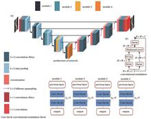
ObjectiveComputed tomography (CT) is an imaging technique that employs X-ray transmission and multi-angle projection to reconstruct the internal structure of an object. Meanwhile, it is commonly adopted in medical diagnosis and industrial non-destructive testing due to its non-invasive and intuitive characteristics. Parallel translational computed tomography (PTCT) acquires projection data by moving a flat panel detector (FPD) and a radiation source in parallel linear motion relative to the detection object. This method has promising applications in industrial inspection. Due to the limitations of the inspection environment and the structure of the inspection system, there are scenarios where it is difficult to realize multi-segment PTCT scanning and imaging, and only single-segment PTCT scanning and imaging can be performed. Since the single-segment PTCT can only obtain the equivalent projection data at a limited angle, its reconstruction problem belongs to limited-angle CT reconstruction. Images reconstructed by traditional algorithms will suffer from serious artifacts. Deep learning-based limited-angle CT image reconstruction has yielded remarkable results, among which model-based data-driven methods have caught much attention. However, such deep networks with CNNs as the main structure tend to focus on the local neighborhood information of the image and ignore the non-local features. Additionally, research on iterative algorithms shows that non-local features can improve detail preservation, which is important for limited-angle CT reconstruction.MethodsTo address the limited-angle artifact in PTCT image reconstruction, we propose a deep iterative unfolding method (STICA-Net, Fig. 3) that learns local and non-local regular terms. The method unfolds a gradient descent algorithm with a fixed number of iterations to a neural network and utilizes convolutional modules with the coordinate attention (CA) mechanism and Swin-Transformer modules deployed as iterative modules in alternating cascades to form an end-to-end deep reconstruction network. The convolution module learns local regularization, in which CA is leveraged to reduce image smoothing. The Swin-Transformer module learns non-local regularization to improve the network's ability to restore image details. Among neighboring modules, iterative connection (IC) is adopted to enhance the model's ability to extract deeper features and improve the efficiency of each iteration. The employed experimental comparison methods are FBP, SIRT, SwinIR, FISTA-Net, and LEARN. The quality of the reconstructed image is comprehensively evaluated by utilizing three sets of quantitative indicators of root mean square error (RMSE), peak signal-to-noise ratio (PSNR), and structural similarity index (SSIM). Meanwhile, comparison experiments are conducted on both simulated and real datasets to verify the feasibility of the proposed method. Additionally, we perform ablation experiments to confirm the effectiveness of each component of the network.Results and DiscussionsWe present the results of a contrast experiment of 90° limited-angle rotational scanning CT using the simulation data 2DeteCT dataset. The results demonstrate the effectiveness of the STICA-Net method for limited-angle reconstruction (Fig. 7). It is noted that PTCT image reconstruction is a limited-angle problem. To verify STICA-Net's effectiveness in PTCT limited-angle reconstruction, we employ the same dataset to generate projection data with an equivalent scanning angle of 90° via PTCT scanning, and then compare different methods. The results of both subjective image evaluation (Fig. 8) and quantitative evaluation index (Table 2) show that STICA-Net can solve the limited-angle problem of PTCT and achieve high-quality image reconstruction. By building the PTCT experimental platform (Fig. 6), the actual dataset of carbon fiber composite core wire (ACCC) is obtained. The two example results (Fig. 11) of the ACCC dataset indicate that the reconstructed images of the traditional method still contain a significant number of artifacts in the absence of large-angle data. However, the artifacts in the reconstructed images of FISTA-Net and LEARN have been significantly reduced. Although FISTA-Net produces better reconstruction results than LEARN, the details are still somewhat blurred. Compared with the suboptimal SwinIR, the PSNR of STICA-Net increases by 4.72% and 5.53%, the SSIM rises by 2.88% and 1.59%, and the RMSE decreases by 15.94% and 19.32% respectively. Meanwhile, ablation experiments verify the effectiveness of different network structures in PTCT limited-angle reconstruction. Figure 10 demonstrates clear improvement in the numerical values of each index as network structures are added incrementally.ConclusionsTo deal with the difficulty of PTCT image reconstruction, we theoretically conclude that PTCT image reconstruction is a limited-angle problem by building a PTCT geometric model, and then propose the STICA-Net model. Ablation experiments confirm the effectiveness of each model component in improving the reconstructed image. Compared to the contrast algorithm, the proposed method significantly improves image quality and yields the best quantitative evaluation indicators across different data types. Additionally, comprehensive results demonstrate that the proposed method outperforms the contrast algorithm in terms of PTCT limited-angle artifact suppression and detail recovery, and high-quality image reconstruction can be achieved. This is beneficial for promoting the in-service detection application of PTCT. However, the method's limitation is that although the ablation experiments demonstrate that the inclusion of the Swin-Transformer structure enhances image results, more memory is needed to store weights and intermediate features, which restricts the utilization of higher-resolution images in our study. In the future, the network module will be further improved to make the network more lightweight. ObjectiveComputed tomography (CT) is an imaging technique that employs X-ray transmission and multi-angle projection to reconstruct the internal structure of an object. Meanwhile, it is commonly adopted in medical diagnosis and industrial non-destructive testing due to its non-invasive and intuitive characteristics. Parallel translational computed tomography (PTCT) acquires projection data by moving a flat panel detector (FPD) and a radiation source in parallel linear motion relative to the detection object. This method has promising applications in industrial inspection. Due to the limitations of the inspection environment and the structure of the inspection system, there are scenarios where it is difficult to realize multi-segment PTCT scanning and imaging, and only single-segment PTCT scanning and imaging can be performed. Since the single-segment PTCT can only obtain the equivalent projection data at a limited angle, its reconstruction problem belongs to limited-angle CT reconstruction. Images reconstructed by traditional algorithms will suffer from serious artifacts. Deep learning-based limited-angle CT image reconstruction has yielded remarkable results, among which model-based data-driven methods have caught much attention. However, such deep networks with CNNs as the main structure tend to focus on the local neighborhood information of the image and ignore the non-local features. Additionally, research on iterative algorithms shows that non-local features can improve detail preservation, which is important for limited-angle CT reconstruction.MethodsTo address the limited-angle artifact in PTCT image reconstruction, we propose a deep iterative unfolding method (STICA-Net, Fig. 3) that learns local and non-local regular terms. The method unfolds a gradient descent algorithm with a fixed number of iterations to a neural network and utilizes convolutional modules with the coordinate attention (CA) mechanism and Swin-Transformer modules deployed as iterative modules in alternating cascades to form an end-to-end deep reconstruction network. The convolution module learns local regularization, in which CA is leveraged to reduce image smoothing. The Swin-Transformer module learns non-local regularization to improve the network's ability to restore image details. Among neighboring modules, iterative connection (IC) is adopted to enhance the model's ability to extract deeper features and improve the efficiency of each iteration. The employed experimental comparison methods are FBP, SIRT, SwinIR, FISTA-Net, and LEARN. The quality of the reconstructed image is comprehensively evaluated by utilizing three sets of quantitative indicators of root mean square error (RMSE), peak signal-to-noise ratio (PSNR), and structural similarity index (SSIM). Meanwhile, comparison experiments are conducted on both simulated and real datasets to verify the feasibility of the proposed method. Additionally, we perform ablation experiments to confirm the effectiveness of each component of the network.Results and DiscussionsWe present the results of a contrast experiment of 90° limited-angle rotational scanning CT using the simulation data 2DeteCT dataset. The results demonstrate the effectiveness of the STICA-Net method for limited-angle reconstruction (Fig. 7). It is noted that PTCT image reconstruction is a limited-angle problem. To verify STICA-Net's effectiveness in PTCT limited-angle reconstruction, we employ the same dataset to generate projection data with an equivalent scanning angle of 90° via PTCT scanning, and then compare different methods. The results of both subjective image evaluation (Fig. 8) and quantitative evaluation index (Table 2) show that STICA-Net can solve the limited-angle problem of PTCT and achieve high-quality image reconstruction. By building the PTCT experimental platform (Fig. 6), the actual dataset of carbon fiber composite core wire (ACCC) is obtained. The two example results (Fig. 11) of the ACCC dataset indicate that the reconstructed images of the traditional method still contain a significant number of artifacts in the absence of large-angle data. However, the artifacts in the reconstructed images of FISTA-Net and LEARN have been significantly reduced. Although FISTA-Net produces better reconstruction results than LEARN, the details are still somewhat blurred. Compared with the suboptimal SwinIR, the PSNR of STICA-Net increases by 4.72% and 5.53%, the SSIM rises by 2.88% and 1.59%, and the RMSE decreases by 15.94% and 19.32% respectively. Meanwhile, ablation experiments verify the effectiveness of different network structures in PTCT limited-angle reconstruction. Figure 10 demonstrates clear improvement in the numerical values of each index as network structures are added incrementally.ConclusionsTo deal with the difficulty of PTCT image reconstruction, we theoretically conclude that PTCT image reconstruction is a limited-angle problem by building a PTCT geometric model, and then propose the STICA-Net model. Ablation experiments confirm the effectiveness of each model component in improving the reconstructed image. Compared to the contrast algorithm, the proposed method significantly improves image quality and yields the best quantitative evaluation indicators across different data types. Additionally, comprehensive results demonstrate that the proposed method outperforms the contrast algorithm in terms of PTCT limited-angle artifact suppression and detail recovery, and high-quality image reconstruction can be achieved. This is beneficial for promoting the in-service detection application of PTCT. However, the method's limitation is that although the ablation experiments demonstrate that the inclusion of the Swin-Transformer structure enhances image results, more memory is needed to store weights and intermediate features, which restricts the utilization of higher-resolution images in our study. In the future, the network module will be further improved to make the network more lightweight.
Acta Optica Sinica
- Publication Date: Apr. 25, 2024
- Vol. 44, Issue 8, 0834001 (2024)

Source Blur Elimination in Micro-CT Using Self-Attention-Based U-Net
Chuanjiang Liu, Ao Wang, Genyuan Zhang, Wei Yuan, and Fenglin Liu
ObjectiveSpatial resolution of X-ray imaging systems is crucial for microstructural object studies due to the small size of the subjects. Specifically, the focal spot size of the X-ray source is a main factor affecting the spatial resolution of micro-computed tomography (micro-CT), which will produce penumbra blur on detectors and thus blur the reconstructed images and reduce spatial resolution. Meanwhile, reducing the focal spot size by decreasing the X-ray tube power is a straightforward solution, but will prolong the scan duration. Therefore, we aim to develop a deep learning-based strategy by learning the inverse finite focal spot model to mitigate the penumbra blur for obtaining CT images with high spatial resolution even in the case of a non-ideal X-ray source.MethodsFirst, we derive the finite focal spot model that builds a relationship from the ideal point source projection to the finite focal spot projection. Based on the derived model, we numerically compute a paired projection dataset. Second, we utilize the neural network U-net and an attention mechanism module of convolution modulation block to build a self-attention mechanism-based U-net (SU-net) and thus learn the inverse finite focal spot model. The goal is to estimate the ideal point source projection from the actual non-ideal focal spot projection. SU-net (Fig. 1) which introduces convolution modulation blocks into the contracting path of the U-net is proposed to boost the U-net property. Finally, the standard filtered back-projection (FBP) is employed for reconstruction using the estimated ideal point projection.Results and DiscussionsSimulation experiments are performed by the public dataset 2DeteCT to verify the effectiveness of the SU-net, which consists of a wide variety of dried fruits, nuts, and different types of rocks. Two groups of results are randomly selected in the test dataset for visualization (Fig. 2) and quantitative indicators are tested on the whole test dataset (Fig. 3). The results show that our proposed SU-net can estimate the ideal point source projection from the non-ideal focal spot projection. To verify the robustness of the SU-net, we test it with data outside of the simulation experimental dataset (Fig. 4), and the results show that it has better generalization than the end-to-end enhanced super resolution generative adversarial network (ESRGAN). Meanwhile, the ablation experiment is conducted with the same dataset and experimental parameters as the simulation experiment to confirm the validity of the added convolutional modulation module (CM) and gradient deviation loss, with quantitative indicators measured (Table 1). The results show that both the CM module and gradient deviation loss added by us can improve the network performance. Practical experiments are carried out to evaluate the effectiveness of the SU-net algorithm on real data (Fig. 5). Since it is difficult to obtain label data in the actual experiment, we select three evaluation indicators that do not require label data (Table 2), including PIQE (perception-based image quality evaluator), NIQE (natural image quality), and image sharpness evaluation function DCT (discrete cosine transform). The results show that our proposed SU-net algorithm achieves the optimal results compared with the comparison methods.ConclusionsIn micro-CT imaging, the focal spot size of the actual X-ray source is limited, and under the relatively large focal spot size, the projected image will be blurred, and the reconstruction of the measured projection directly using the CT algorithm based on the point source model will cause the image to be blurred. We propose a U-net based on the self-attention mechanism to estimate the ideal point source projection from the actual measured non-ideal focal spot projection. Meanwhile, we establish a training dataset according to the relationship between the non-ideal focal spot projection and the ideal point source projection to optimize the network. Simulation and practical experiments show that this method can effectively estimate clear projection from blurred projection. The advantage of the proposed method is that we can construct a dataset by the relationship between the finite focal spot projection model and the ideal point source projection model, without collecting data pairs composed of non-ideal focal spot projection and ideal point source projection, which greatly reduces the difficulty of constructing datasets. Secondly, the proposed network directly based on the relationship between the finite focal spot projection model and the ideal point source projection model has strong interpretability, which means the inverse relationship from the finite focal spot model to the ideal point source model is learned through the network. Therefore, this method has better generalization than end-to-end ESRGAN, especially for CT images with high fidelity of image details. Our limitation is that the training is conducted for a specific focal spot size and a specific scanning geometry without considering the influence of noise. Subsequent studies will train networks with different focal spot sizes and geometric parameters and consider situations with noise. ObjectiveSpatial resolution of X-ray imaging systems is crucial for microstructural object studies due to the small size of the subjects. Specifically, the focal spot size of the X-ray source is a main factor affecting the spatial resolution of micro-computed tomography (micro-CT), which will produce penumbra blur on detectors and thus blur the reconstructed images and reduce spatial resolution. Meanwhile, reducing the focal spot size by decreasing the X-ray tube power is a straightforward solution, but will prolong the scan duration. Therefore, we aim to develop a deep learning-based strategy by learning the inverse finite focal spot model to mitigate the penumbra blur for obtaining CT images with high spatial resolution even in the case of a non-ideal X-ray source.MethodsFirst, we derive the finite focal spot model that builds a relationship from the ideal point source projection to the finite focal spot projection. Based on the derived model, we numerically compute a paired projection dataset. Second, we utilize the neural network U-net and an attention mechanism module of convolution modulation block to build a self-attention mechanism-based U-net (SU-net) and thus learn the inverse finite focal spot model. The goal is to estimate the ideal point source projection from the actual non-ideal focal spot projection. SU-net (Fig. 1) which introduces convolution modulation blocks into the contracting path of the U-net is proposed to boost the U-net property. Finally, the standard filtered back-projection (FBP) is employed for reconstruction using the estimated ideal point projection.Results and DiscussionsSimulation experiments are performed by the public dataset 2DeteCT to verify the effectiveness of the SU-net, which consists of a wide variety of dried fruits, nuts, and different types of rocks. Two groups of results are randomly selected in the test dataset for visualization (Fig. 2) and quantitative indicators are tested on the whole test dataset (Fig. 3). The results show that our proposed SU-net can estimate the ideal point source projection from the non-ideal focal spot projection. To verify the robustness of the SU-net, we test it with data outside of the simulation experimental dataset (Fig. 4), and the results show that it has better generalization than the end-to-end enhanced super resolution generative adversarial network (ESRGAN). Meanwhile, the ablation experiment is conducted with the same dataset and experimental parameters as the simulation experiment to confirm the validity of the added convolutional modulation module (CM) and gradient deviation loss, with quantitative indicators measured (Table 1). The results show that both the CM module and gradient deviation loss added by us can improve the network performance. Practical experiments are carried out to evaluate the effectiveness of the SU-net algorithm on real data (Fig. 5). Since it is difficult to obtain label data in the actual experiment, we select three evaluation indicators that do not require label data (Table 2), including PIQE (perception-based image quality evaluator), NIQE (natural image quality), and image sharpness evaluation function DCT (discrete cosine transform). The results show that our proposed SU-net algorithm achieves the optimal results compared with the comparison methods.ConclusionsIn micro-CT imaging, the focal spot size of the actual X-ray source is limited, and under the relatively large focal spot size, the projected image will be blurred, and the reconstruction of the measured projection directly using the CT algorithm based on the point source model will cause the image to be blurred. We propose a U-net based on the self-attention mechanism to estimate the ideal point source projection from the actual measured non-ideal focal spot projection. Meanwhile, we establish a training dataset according to the relationship between the non-ideal focal spot projection and the ideal point source projection to optimize the network. Simulation and practical experiments show that this method can effectively estimate clear projection from blurred projection. The advantage of the proposed method is that we can construct a dataset by the relationship between the finite focal spot projection model and the ideal point source projection model, without collecting data pairs composed of non-ideal focal spot projection and ideal point source projection, which greatly reduces the difficulty of constructing datasets. Secondly, the proposed network directly based on the relationship between the finite focal spot projection model and the ideal point source projection model has strong interpretability, which means the inverse relationship from the finite focal spot model to the ideal point source model is learned through the network. Therefore, this method has better generalization than end-to-end ESRGAN, especially for CT images with high fidelity of image details. Our limitation is that the training is conducted for a specific focal spot size and a specific scanning geometry without considering the influence of noise. Subsequent studies will train networks with different focal spot sizes and geometric parameters and consider situations with noise.
Acta Optica Sinica
- Publication Date: Apr. 10, 2024
- Vol. 44, Issue 7, 0734002 (2024)

Cosmic Ray Rejection in Small Angle X-Ray Scattering
Jin Zhao, Chenglong Wang, and Hong Yu
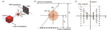
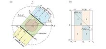
ObjectiveSmall angle X-ray scattering (SAXS) is a powerful tool to measure structural features on the order of 1-100 nm. Due to high measurement accuracy and strong penetrability, SAXS attracts much attention to characterizing the complex three-dimensional (3D) structure information of periodic nanostructures in integrated circuit (IC) and has been successfully applied to high aspect ratio (HAR) structures, such as 3D-NAND and DRAM. SAXS for IC inline metrology is mostly based on compact X-ray sources. Limited by the brightness of compact X-ray sources, SAXS measurement requires a long exposure time to improve the signal-to-noise (SNR) of SAXS signals. Since the integration effect of long exposure time, numerous cosmic rays are inevitably introduced in the SAXS measurement pattern. As a typical kind of noise that is not correlated with SAXS signals, cosmic rays appear in SAXS patterns randomly and cause signal distortion, which has a negative effect on nanostructure information extraction. However, for lack of making full use of the signal's periodicity information, present cosmic ray rejection algorithms cannot accurately identify and remove the cosmic rays that have a real influence on SAXS signals in the measurement pattern. A new cosmic ray rejection method is needed for SAXS measurement patterns of periodic nanostructures, which will help improve the SNR of SAXS patterns and the performance of nanostructure information extraction.MethodsWe propose a cosmic ray rejection method for the SAXS measurement pattern of periodic nanostructure. First, a pattern sequence including many short exposure SAXS measurement patterns of periodic nanostructure samples is generated in the same measurement conditions. Then, the coordinates of the periodic scattering signals are calculated by taking the periodic information of the nanostructure as physical prior, and cosmic rays existing in the effective signal area for each diffraction order in each scattering pattern are identified. After removing the abnormal frames influenced by cosmic rays from the pattern sequence, the SAXS measurement pattern after cosmic ray rejection is obtained by summing the remaining frames of the pattern sequence. The pattern sequence including 500 short exposure SAXS measurement patterns of periodic nanostructure samples is used to evaluate the performance of the proposed method. The precision, miss rate, and false alarm rate of cosmic ray detection results of the pattern sequence are calculated. Meanwhile, two existing methods for cosmic ray rejection of Laplacian edge detection and multi-frame median pixel rejection are selected as the comparison method, and the SAXS measurement pattern sequence is removed from cosmic rays by adopting the two comparison methods and the proposed method. The mean square error (MSE), peak signal-to-noise ratio (PSNR), and structural similarity (SSIM) of the pattern sequence before and after cosmic ray rejection by three methods are calculated respectively. Since the influence of cosmic rays and Poisson noise on the SAXS measurement pattern is relative to the exposure time, the competitive relationships between two kinds of noise and cosmic ray rejection performances of the proposed method in different exposure times are analyzed. This is realized by calculating the relationship of PSNR of the pattern sequence before and after denoising by three methods respectively, and the number of frames included in the sequence.Results and DiscussionsAccording to the confusion matrix calculated based on the cosmic ray detection results of the pattern sequence including 500 short exposure SAXS measurement patterns (Fig. 4), the precision, miss rate, and false alarm rate are 87.67%, 4.93%, and 5.18%, respectively. Compared with the two comparison methods, the pattern sequence denoised by this method has the best cosmic ray rejection effect, and the MSE, PSNR, and SSIM of the method are all optimal (Fig. 5 and Table 1). Especially, PSNR of the pattern sequence increases by 5.55 dB after removing cosmic rays by this method. When the number of frames included in the pattern sequence is low and equivalent to exposure time, Poisson noise is dominant and the PSNR of the pattern sequence is so low that we need more exposure time. When the number of frames increases to about 200, cosmic rays seriously restrict the upper limit of the PSNR of the scattering pattern. However, the PSNR of the pattern sequence after denoising by this method still increases with the rising number of frames, and the growth rate of the PSNR is significantly higher than comparison methods (Fig. 6).ConclusionsWe propose a method for cosmic ray rejection in the SAXS measurement pattern of periodic nanostructures. The pattern sequence including 500 short exposure SAXS measurement patterns of periodic nanostructure samples is simulated and removed from cosmic rays by this method. According to the cosmic ray detection results, the miss rate and false alarm rate are both only about 5%, which proves that the proposed method has a sound detection effect on cosmic rays for the single frame scattering pattern. Meanwhile, the PSNR of the pattern sequence increases by 5.55 dB after removing cosmic rays by the proposed method. The PSNR gain greatly improves the extraction reliability and accuracy of the periodic nanostructure information. By analyzing the competitive relationship between Poisson noise and cosmic rays and evaluating the cosmic ray rejection performance of the proposed method in different exposure time, we find that this method can break the upper limit of PSNR caused by cosmic rays and improve PSNR of scattering pattern continuously. This proves that this method can obtain excellent PSNR gain in the long exposure integration condition. In principle, the proposed method provides a reliable cosmic ray rejection scheme for SAXS measurement patterns of periodic nanostructures, improving the detection SNR of SAXS patterns effectively. This method features a simple principle and fast operation and thus has practical significance to improve the inline metrology performance of SAXS. ObjectiveSmall angle X-ray scattering (SAXS) is a powerful tool to measure structural features on the order of 1-100 nm. Due to high measurement accuracy and strong penetrability, SAXS attracts much attention to characterizing the complex three-dimensional (3D) structure information of periodic nanostructures in integrated circuit (IC) and has been successfully applied to high aspect ratio (HAR) structures, such as 3D-NAND and DRAM. SAXS for IC inline metrology is mostly based on compact X-ray sources. Limited by the brightness of compact X-ray sources, SAXS measurement requires a long exposure time to improve the signal-to-noise (SNR) of SAXS signals. Since the integration effect of long exposure time, numerous cosmic rays are inevitably introduced in the SAXS measurement pattern. As a typical kind of noise that is not correlated with SAXS signals, cosmic rays appear in SAXS patterns randomly and cause signal distortion, which has a negative effect on nanostructure information extraction. However, for lack of making full use of the signal's periodicity information, present cosmic ray rejection algorithms cannot accurately identify and remove the cosmic rays that have a real influence on SAXS signals in the measurement pattern. A new cosmic ray rejection method is needed for SAXS measurement patterns of periodic nanostructures, which will help improve the SNR of SAXS patterns and the performance of nanostructure information extraction.MethodsWe propose a cosmic ray rejection method for the SAXS measurement pattern of periodic nanostructure. First, a pattern sequence including many short exposure SAXS measurement patterns of periodic nanostructure samples is generated in the same measurement conditions. Then, the coordinates of the periodic scattering signals are calculated by taking the periodic information of the nanostructure as physical prior, and cosmic rays existing in the effective signal area for each diffraction order in each scattering pattern are identified. After removing the abnormal frames influenced by cosmic rays from the pattern sequence, the SAXS measurement pattern after cosmic ray rejection is obtained by summing the remaining frames of the pattern sequence. The pattern sequence including 500 short exposure SAXS measurement patterns of periodic nanostructure samples is used to evaluate the performance of the proposed method. The precision, miss rate, and false alarm rate of cosmic ray detection results of the pattern sequence are calculated. Meanwhile, two existing methods for cosmic ray rejection of Laplacian edge detection and multi-frame median pixel rejection are selected as the comparison method, and the SAXS measurement pattern sequence is removed from cosmic rays by adopting the two comparison methods and the proposed method. The mean square error (MSE), peak signal-to-noise ratio (PSNR), and structural similarity (SSIM) of the pattern sequence before and after cosmic ray rejection by three methods are calculated respectively. Since the influence of cosmic rays and Poisson noise on the SAXS measurement pattern is relative to the exposure time, the competitive relationships between two kinds of noise and cosmic ray rejection performances of the proposed method in different exposure times are analyzed. This is realized by calculating the relationship of PSNR of the pattern sequence before and after denoising by three methods respectively, and the number of frames included in the sequence.Results and DiscussionsAccording to the confusion matrix calculated based on the cosmic ray detection results of the pattern sequence including 500 short exposure SAXS measurement patterns (Fig. 4), the precision, miss rate, and false alarm rate are 87.67%, 4.93%, and 5.18%, respectively. Compared with the two comparison methods, the pattern sequence denoised by this method has the best cosmic ray rejection effect, and the MSE, PSNR, and SSIM of the method are all optimal (Fig. 5 and Table 1). Especially, PSNR of the pattern sequence increases by 5.55 dB after removing cosmic rays by this method. When the number of frames included in the pattern sequence is low and equivalent to exposure time, Poisson noise is dominant and the PSNR of the pattern sequence is so low that we need more exposure time. When the number of frames increases to about 200, cosmic rays seriously restrict the upper limit of the PSNR of the scattering pattern. However, the PSNR of the pattern sequence after denoising by this method still increases with the rising number of frames, and the growth rate of the PSNR is significantly higher than comparison methods (Fig. 6).ConclusionsWe propose a method for cosmic ray rejection in the SAXS measurement pattern of periodic nanostructures. The pattern sequence including 500 short exposure SAXS measurement patterns of periodic nanostructure samples is simulated and removed from cosmic rays by this method. According to the cosmic ray detection results, the miss rate and false alarm rate are both only about 5%, which proves that the proposed method has a sound detection effect on cosmic rays for the single frame scattering pattern. Meanwhile, the PSNR of the pattern sequence increases by 5.55 dB after removing cosmic rays by the proposed method. The PSNR gain greatly improves the extraction reliability and accuracy of the periodic nanostructure information. By analyzing the competitive relationship between Poisson noise and cosmic rays and evaluating the cosmic ray rejection performance of the proposed method in different exposure time, we find that this method can break the upper limit of PSNR caused by cosmic rays and improve PSNR of scattering pattern continuously. This proves that this method can obtain excellent PSNR gain in the long exposure integration condition. In principle, the proposed method provides a reliable cosmic ray rejection scheme for SAXS measurement patterns of periodic nanostructures, improving the detection SNR of SAXS patterns effectively. This method features a simple principle and fast operation and thus has practical significance to improve the inline metrology performance of SAXS.
Acta Optica Sinica
- Publication Date: Apr. 10, 2024
- Vol. 44, Issue 7, 0734001 (2024)
Simulation of Polarimetric Photoelectric Process in X-Ray Polarization Detector
Renzhou Zheng, Pengfei Qiang, Lizhi Sheng, and Yongqing Yan
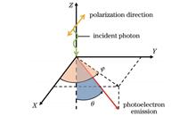
ObjectiveX-ray polarization detection is an important means to study the astrophysical properties of intense X-ray sources such as black holes, pulsars, and related gamma-ray bursts. The development of X-ray polarization detectors with excellent performance is the technical basis for related research. Early X-ray polarization detectors were mainly Thomson scattering polarimeters and Bragg polarimeters. However, due to the low modulation factor and narrow detection energy range, the ideal polarization measurement results were not obtained. In 2001, Costa et al. proposed a new way of X-ray polarization detection using the photoelectric effect, in which the X-ray polarization information was obtained by imaging the photoelectron track produced by X-ray photons through a gas detector. The polarimetric photoelectric process is the key physical process for the detector to realize polarization detection. It is of great significance to clarify the photon-gas interaction process and the distribution law of emitted photoelectrons for further understanding the working mechanism of the detector. The polarimetric photoelectric process is an important research content in the development of this type of X-ray polarization detector. Different types of gases have various properties, which will affect the particle transport in the polarimetric photoelectric process and further leads to different detection efficiencies. Therefore, it is necessary to simulate the polarimetric photoelectric process under different conditions. This can provide a theoretical basis and data support for the structure design of X-ray polarization detectors.MethodsWe simulate the polarimetric photoelectric process of 2-10 keV linearly polarized X-ray photons in several commonly used working gases by the Monte Carlo code Geant4. The selected working gas combinations include He+C3H8, Ne+CF4, Ne+DME, Ar+CH4, Ar+CO2, Xe+CO2, CF4+C4H10, and DME+CO2. The response relationship of the emission position and azimuthal angle distribution of photoelectron with the polarization direction and energy of the incident photon is discussed. Moreover, the effects of gas thickness, gas component, gas ratio, and photon energy on the detection efficiency are analyzed.Results and DiscussionsFirst, the response relationship of the emission position and azimuthal angle distribution of the photoelectron with the polarization direction and energy of the incident photon is clarified. The emission direction distribution probability of the photoelectron is the largest in the polarization direction of the incident photon, and the azimuthal angle distribution can be approximated as a cosine squared function. With the increase in photon energy, the counts of photoelectrons at each angle decrease in different degrees, but all of them show a statistical law that the maximum values occur when the azimuthal angle is 0 or π (-π) (Fig. 6). Moreover, the effects of gas thickness, gas component, gas ratio, and photon energy on the detection efficiency are revealed and quantified. For 2 keV photons entering into 90%Ne+10%DME gas mixture, when the gas thickness is small, the detection efficiency increases rapidly with the increase in gas thickness, from less than 0.1 at 0.1 cm to 0.64 at 1 cm (Fig. 7). When the gas thickness increases to 3 cm, the detection efficiency is greater than 0.9. Then, with the increase in gas thickness, the detection efficiency gradually approaches 1. For the CF4+C4H10, Ne+CF4, Ne+DME, DME+CO2, and He+C3H8, the detection efficiency decreases with the increase in photon energy, and the large average atomic number of gas can lead to a high detection efficiency (Fig. 8). While for the Xe+CO2, Ar+CO2, and Ar+CH4, when the photon energy is greater than the binding energy of certain shell electrons of Xe or Ar atoms, the detection efficiency will be improved to a certain extent because the corresponding shell electrons begin to be ejected. In addition to the Ar+CO2 which is affected by the electron emission in K-shell, the detection efficiency in each energy range can be effectively improved by increasing the proportion of gas with high atomic number (Fig. 9).ConclusionsWe simulate the polarimetric photoelectric process of 2-10 keV linearly polarized X-ray photons in several commonly used working gases by the Monte Carlo code Geant4. The response relationship of the emission position and azimuthal angle distribution of the photoelectron with the polarization direction and energy of the incident photon is clarified. The emission direction distribution probability of the photoelectron is the largest on the polarization direction of the incident photon, and the azimuthal angle distribution can be approximated as a cosine squared function. With the increase in photon energy, the counts of photoelectrons at each angle decrease in different degrees, but all of them show a statistical law that the maximum values occur when the azimuthal angle is 0 or π (-π). Moreover, the effects of gas thickness, gas component, gas ratio, and photon energy on the detection efficiency are revealed and quantified. The larger gas thickness and larger average atomic number can lead to higher detection efficiency. In addition, the increase in photon energy can result in a decrease in detection efficiency. However, for the working gases composed of Xe or Ar, when the photon energy is greater than the binding energy of a certain shell electron, the detection efficiency will be improved to a certain extent because the corresponding shell electrons begin to be ejected. The results in this paper can provide some theoretical basis and data support for the structure design of X-ray polarization detectors. In the actual selection of working gases, the drift properties of electrons in gases, the effect of photoelectron drift and diffusion on track thickness and length, and the reconstruction efficiency of the track reconstruction algorithm should also be considered. ObjectiveX-ray polarization detection is an important means to study the astrophysical properties of intense X-ray sources such as black holes, pulsars, and related gamma-ray bursts. The development of X-ray polarization detectors with excellent performance is the technical basis for related research. Early X-ray polarization detectors were mainly Thomson scattering polarimeters and Bragg polarimeters. However, due to the low modulation factor and narrow detection energy range, the ideal polarization measurement results were not obtained. In 2001, Costa et al. proposed a new way of X-ray polarization detection using the photoelectric effect, in which the X-ray polarization information was obtained by imaging the photoelectron track produced by X-ray photons through a gas detector. The polarimetric photoelectric process is the key physical process for the detector to realize polarization detection. It is of great significance to clarify the photon-gas interaction process and the distribution law of emitted photoelectrons for further understanding the working mechanism of the detector. The polarimetric photoelectric process is an important research content in the development of this type of X-ray polarization detector. Different types of gases have various properties, which will affect the particle transport in the polarimetric photoelectric process and further leads to different detection efficiencies. Therefore, it is necessary to simulate the polarimetric photoelectric process under different conditions. This can provide a theoretical basis and data support for the structure design of X-ray polarization detectors.MethodsWe simulate the polarimetric photoelectric process of 2-10 keV linearly polarized X-ray photons in several commonly used working gases by the Monte Carlo code Geant4. The selected working gas combinations include He+C3H8, Ne+CF4, Ne+DME, Ar+CH4, Ar+CO2, Xe+CO2, CF4+C4H10, and DME+CO2. The response relationship of the emission position and azimuthal angle distribution of photoelectron with the polarization direction and energy of the incident photon is discussed. Moreover, the effects of gas thickness, gas component, gas ratio, and photon energy on the detection efficiency are analyzed.Results and DiscussionsFirst, the response relationship of the emission position and azimuthal angle distribution of the photoelectron with the polarization direction and energy of the incident photon is clarified. The emission direction distribution probability of the photoelectron is the largest in the polarization direction of the incident photon, and the azimuthal angle distribution can be approximated as a cosine squared function. With the increase in photon energy, the counts of photoelectrons at each angle decrease in different degrees, but all of them show a statistical law that the maximum values occur when the azimuthal angle is 0 or π (-π) (Fig. 6). Moreover, the effects of gas thickness, gas component, gas ratio, and photon energy on the detection efficiency are revealed and quantified. For 2 keV photons entering into 90%Ne+10%DME gas mixture, when the gas thickness is small, the detection efficiency increases rapidly with the increase in gas thickness, from less than 0.1 at 0.1 cm to 0.64 at 1 cm (Fig. 7). When the gas thickness increases to 3 cm, the detection efficiency is greater than 0.9. Then, with the increase in gas thickness, the detection efficiency gradually approaches 1. For the CF4+C4H10, Ne+CF4, Ne+DME, DME+CO2, and He+C3H8, the detection efficiency decreases with the increase in photon energy, and the large average atomic number of gas can lead to a high detection efficiency (Fig. 8). While for the Xe+CO2, Ar+CO2, and Ar+CH4, when the photon energy is greater than the binding energy of certain shell electrons of Xe or Ar atoms, the detection efficiency will be improved to a certain extent because the corresponding shell electrons begin to be ejected. In addition to the Ar+CO2 which is affected by the electron emission in K-shell, the detection efficiency in each energy range can be effectively improved by increasing the proportion of gas with high atomic number (Fig. 9).ConclusionsWe simulate the polarimetric photoelectric process of 2-10 keV linearly polarized X-ray photons in several commonly used working gases by the Monte Carlo code Geant4. The response relationship of the emission position and azimuthal angle distribution of the photoelectron with the polarization direction and energy of the incident photon is clarified. The emission direction distribution probability of the photoelectron is the largest on the polarization direction of the incident photon, and the azimuthal angle distribution can be approximated as a cosine squared function. With the increase in photon energy, the counts of photoelectrons at each angle decrease in different degrees, but all of them show a statistical law that the maximum values occur when the azimuthal angle is 0 or π (-π). Moreover, the effects of gas thickness, gas component, gas ratio, and photon energy on the detection efficiency are revealed and quantified. The larger gas thickness and larger average atomic number can lead to higher detection efficiency. In addition, the increase in photon energy can result in a decrease in detection efficiency. However, for the working gases composed of Xe or Ar, when the photon energy is greater than the binding energy of a certain shell electron, the detection efficiency will be improved to a certain extent because the corresponding shell electrons begin to be ejected. The results in this paper can provide some theoretical basis and data support for the structure design of X-ray polarization detectors. In the actual selection of working gases, the drift properties of electrons in gases, the effect of photoelectron drift and diffusion on track thickness and length, and the reconstruction efficiency of the track reconstruction algorithm should also be considered.
Acta Optica Sinica
- Publication Date: Feb. 10, 2024
- Vol. 44, Issue 3, 0334003 (2024)
Neutron Displaced Computed Tomography Scanning Imaging Method Considering Calibration Error of Center of Rotation
Qiang Lin, Zeming Ma, Bin Liu, Wenjian Wang, Haohao Ding, and Min Yang
ObjectiveNeutron displaced CT (computed tomography) scanning is an effective tomography detection method for large-sized samples, but the truncated projection data leads to significant calibration errors of the center of rotation (COR) of the turntable in the CT system, seriously affecting imaging quality. We consider the COR calibration error during the design of the neutron displaced CT scanning imaging method. A COR calibration algorithm of the turntable under the displaced CT scanning is designed. Then, the symmetric complementary data (SCD) reconstruction algorithm and the projection data preprocessing (PDP) reconstruction algorithm are established. The sensitivities of the reconstruction accuracy of the two reconstruction algorithms to the COR calibration error are discussed. We hope that the proposedCOR calibration algorithm and the reconstruction algorithm under the neutron displaced CT scanning mode can lay a theoretical foundation for solving the neutron CT imaging problem of large-sized samples.MethodsA preciseCOR calibration method under the neutron displaced CT scanning mode is established. The calibration algorithm based on the symmetry principle of projection data is designed. Each possible COR position is enumerated, and the variances between the sum of the projection data on the left and the right sides of the COR are calculated. Finally, the COR result is determined by finding the location where the variance has the minimum value. Under the displaced CT scanning mode, the truncation and redundant characteristics of projection data will result in bright circular artifacts in the reconstructed images. We design two reconstruction algorithms to eliminate the bright circular artifacts, namely SCD reconstruction algorithm and PDP reconstruction algorithm. SCD reconstruction algorithm supply the missing projection data under displaced scanning mode by using the principle of symmetric complementary data and then use the filtered back projection (FBP) algorithm to obtain the accurate reconstruction result. PDP reconstruction algorithm utilize the WANG weighting function to process sinogram data. In order to eliminate the bright circular artifacts in the reconstructed image, the redundant projection values are weighted to ensure that the projection data from all directions contribute the same data amount to the reconstruction results. A simulation method of neutron projection noise including Gaussian noise and γ white spot noise is proposed, and a 3D simulation phantom is designed to verify the performance advantages of the proposed COR calibration algorithm and PDP reconstruction algorithm under different COR displaced sizes and projection noise intensities. A neutron displaced CT scanning imaging experiment is conducted based on the reactor neutron source to verify the practicality and stability of the proposed COR calibration algorithm and the reconstruction algorithm.Results and DiscussionsBy using the designed 3D simulation phantom, it can be verified that the proposed COR calibration algorithm has a calibration error of 0.1. After adding Gaussian noise and γ white spot noise to the projection data of the 3D simulation phantom, the noise in the projection image is similar to the actual neutron data. As the noise intensity increases, the COR calculation error of the OAC (opposite angle calibration) algorithm significantly increases, but the COR calibration error of the proposed method does not increase (Table 2). Therefore, it can be proven that the proposed COR calibration algorithm has higher accuracy and stability. When the COR displaced size changes, the calibration error of the COR does not significantly increase. When the COR calibration error reaches two pixels, the reconstruction results of SCD reconstruction algorithm show certain image artifacts, resulting in distortion of the detailed structure in the reconstructed image. Additionally, due to the influence of stitching and misalignment, the reconstructed image also shows certain stripe artifacts (Fig. 8). The reconstruction results obtained by PDP reconstruction algorithm have stronger image detail resolution and higher reconstructed image quality (Fig. 9). When the projection has Gaussian noise and γ white spot noise, the reconstruction results obtained by PDP reconstruction algorithm are also better than those obtained by SCD reconstruction algorithm (Fig. 10). Moreover, PDP reconstruction algorithm can also achieve good reconstruction results when the COR displaced size changes (Fig. 11). Based on the reactor neutron source of China Academy of Engineering Physics, a neutron displaced CT scanning experiment is carried out, and clear internal and external structural details of the sample are obtained. The imaging field of the neutron CT system is expanded by 31.4%.ConclusionsWe design a neutron displaced CT scanning imaging method for large-sized samples and a COR calibration algorithm under the neutron displaced CT scanning based on the symmetry principle of projection data. The proposed COR calibration algorithm has the advantages of high measurement accuracy and strong anti-noise ability. Two neutron displaced CT scanning reconstruction algorithms are developed. SCD reconstruction algorithm is more sensitive to COR calibration errors. A smaller error can lead to stitching and misalignment issues in the supplemented projection data, thereby affecting the quality of image reconstruction. PDP reconstruction algorithm has a strong tolerance for the COR calibration error and can obtain higher reconstructed image quality. The 3D simulation phantom verifies the performance advantages of the proposed calibration algorithm and PDP reconstruction algorithm under different COR displaced sizes and projection noise intensities. In addition, the neutron displaced CT scanning experiment prove that the proposed COR calibration algorithm and PDP reconstruction algorithm have significant engineering practical values, laying a theoretical foundation for solving the problem of neutron CT imaging of large-sized samples. ObjectiveNeutron displaced CT (computed tomography) scanning is an effective tomography detection method for large-sized samples, but the truncated projection data leads to significant calibration errors of the center of rotation (COR) of the turntable in the CT system, seriously affecting imaging quality. We consider the COR calibration error during the design of the neutron displaced CT scanning imaging method. A COR calibration algorithm of the turntable under the displaced CT scanning is designed. Then, the symmetric complementary data (SCD) reconstruction algorithm and the projection data preprocessing (PDP) reconstruction algorithm are established. The sensitivities of the reconstruction accuracy of the two reconstruction algorithms to the COR calibration error are discussed. We hope that the proposedCOR calibration algorithm and the reconstruction algorithm under the neutron displaced CT scanning mode can lay a theoretical foundation for solving the neutron CT imaging problem of large-sized samples.MethodsA preciseCOR calibration method under the neutron displaced CT scanning mode is established. The calibration algorithm based on the symmetry principle of projection data is designed. Each possible COR position is enumerated, and the variances between the sum of the projection data on the left and the right sides of the COR are calculated. Finally, the COR result is determined by finding the location where the variance has the minimum value. Under the displaced CT scanning mode, the truncation and redundant characteristics of projection data will result in bright circular artifacts in the reconstructed images. We design two reconstruction algorithms to eliminate the bright circular artifacts, namely SCD reconstruction algorithm and PDP reconstruction algorithm. SCD reconstruction algorithm supply the missing projection data under displaced scanning mode by using the principle of symmetric complementary data and then use the filtered back projection (FBP) algorithm to obtain the accurate reconstruction result. PDP reconstruction algorithm utilize the WANG weighting function to process sinogram data. In order to eliminate the bright circular artifacts in the reconstructed image, the redundant projection values are weighted to ensure that the projection data from all directions contribute the same data amount to the reconstruction results. A simulation method of neutron projection noise including Gaussian noise and γ white spot noise is proposed, and a 3D simulation phantom is designed to verify the performance advantages of the proposed COR calibration algorithm and PDP reconstruction algorithm under different COR displaced sizes and projection noise intensities. A neutron displaced CT scanning imaging experiment is conducted based on the reactor neutron source to verify the practicality and stability of the proposed COR calibration algorithm and the reconstruction algorithm.Results and DiscussionsBy using the designed 3D simulation phantom, it can be verified that the proposed COR calibration algorithm has a calibration error of 0.1. After adding Gaussian noise and γ white spot noise to the projection data of the 3D simulation phantom, the noise in the projection image is similar to the actual neutron data. As the noise intensity increases, the COR calculation error of the OAC (opposite angle calibration) algorithm significantly increases, but the COR calibration error of the proposed method does not increase (Table 2). Therefore, it can be proven that the proposed COR calibration algorithm has higher accuracy and stability. When the COR displaced size changes, the calibration error of the COR does not significantly increase. When the COR calibration error reaches two pixels, the reconstruction results of SCD reconstruction algorithm show certain image artifacts, resulting in distortion of the detailed structure in the reconstructed image. Additionally, due to the influence of stitching and misalignment, the reconstructed image also shows certain stripe artifacts (Fig. 8). The reconstruction results obtained by PDP reconstruction algorithm have stronger image detail resolution and higher reconstructed image quality (Fig. 9). When the projection has Gaussian noise and γ white spot noise, the reconstruction results obtained by PDP reconstruction algorithm are also better than those obtained by SCD reconstruction algorithm (Fig. 10). Moreover, PDP reconstruction algorithm can also achieve good reconstruction results when the COR displaced size changes (Fig. 11). Based on the reactor neutron source of China Academy of Engineering Physics, a neutron displaced CT scanning experiment is carried out, and clear internal and external structural details of the sample are obtained. The imaging field of the neutron CT system is expanded by 31.4%.ConclusionsWe design a neutron displaced CT scanning imaging method for large-sized samples and a COR calibration algorithm under the neutron displaced CT scanning based on the symmetry principle of projection data. The proposed COR calibration algorithm has the advantages of high measurement accuracy and strong anti-noise ability. Two neutron displaced CT scanning reconstruction algorithms are developed. SCD reconstruction algorithm is more sensitive to COR calibration errors. A smaller error can lead to stitching and misalignment issues in the supplemented projection data, thereby affecting the quality of image reconstruction. PDP reconstruction algorithm has a strong tolerance for the COR calibration error and can obtain higher reconstructed image quality. The 3D simulation phantom verifies the performance advantages of the proposed calibration algorithm and PDP reconstruction algorithm under different COR displaced sizes and projection noise intensities. In addition, the neutron displaced CT scanning experiment prove that the proposed COR calibration algorithm and PDP reconstruction algorithm have significant engineering practical values, laying a theoretical foundation for solving the problem of neutron CT imaging of large-sized samples.
Acta Optica Sinica
- Publication Date: Feb. 10, 2024
- Vol. 44, Issue 3, 0334002 (2024)

Photoelectron Generation and Control of Streak Tubes Based on Geant4-CST Co-Simulation
Yuxiang Liao, Zichen Wang, Lin Tang, Yuming Feng, Xiaoyan Zhao, Diwei Liu, and Kaichun Zhang
ObjectiveIn recent years, inertial confinement fusion (ICF) technology has developed rapidly and exhibited its great potential for applications. In ICF experiments, the pellet will radiate a large amount of X-rays, and the nuclear fusion process can be analyzed by studying the spatio-temporal properties of X-rays. However, the nuclear fusion duration is short (nanosecond-picosecond order), with requirements of high spatial resolution and large dynamic range. However, the commonly applied ultra-rapid diagnostic instruments are more or less defective, among which the optomechanical high-speed camera cannot monitor ultrafast phenomena below the nanosecond order, with sufficient temporal resolution. Electro-optical or magneto-optical shutter high-speed camera makes it difficult to monitor weak signals due to a shutter resulting in incident light loss. Therefore, the study of streak cameras (streak tubes) with ultra-high spatio-temporal and light intensity resolution capabilities is of significance for detecting X-rays in ICF experiments. The anisotropic focusing streak tubes can achieve anisotropic focusing of electron beams by making the temporal-directed focusing system and the spatial-directed focusing system independent of each other. This tube type can not only improve the spatial resolution by increasing the magnification of the streak tube, but also suppress the space charge effect, reduce the aberration in the spatial direction, and improve the dynamic range and temporal resolution of the streak tubes.MethodsAt present, it is difficult for the existing simulation software to completely simulate the whole physical process of streak tubes. Although CST (CST Studio Suite) and other electromagnetic simulation software can suitably reflect the electron transport and the interaction between electrons and electromagnetic fields, less credible results are given for the photoelectron generation process. Therefore, when designing a streak tube, researchers generally need to specially program to calculate the photoelectron distribution of the photocathode based on the Monte Carlo method. However, generally, a programme can only be adopted for one or a few cases, which makes it less portable. Additionally, it is more difficult to verify the results of purely theoretical calculations. Therefore, we employ a high-energy particle simulation software Geant4 (GEometry ANd Tracking) developed by the European Organization for Nuclear Research based on the Monte Carlo method to simulate the photoelectron generation process. Then, based on the simulation results of Geant4, we leverage CST to simulate the subsequent electro-optical system. Finally, the design of an anisotropic focusing streak tube is realized by the software co-simulation.Results and DiscussionsThe high-energy particle simulation software Geant4 is introduced to ultrafast diagnostics, the co-simulation from Geant4 to CST is realized, and an anisotropic focusing streak tube design that encompasses the entire process of photoelectron generation, transmission, focusing, imaging, and interaction between electrons and electromagnetic fields is yielded. Compared with the traditional simulation method, the photoelectron generation process is visualized in this scheme, and the photoelectron distribution is more consistent with the actual experimental situation. Meanwhile, since Geant4 can provide models for the electromagnetic, strong, and weak interaction between substances and particles of different energy to simulate the complete physical process, this scheme can be adapted to a wider range of photoelectron generation situations and is highly portable.ConclusionsBy adopting the co-simulation from Geant4 to CST, an anisotropic focusing streak tube with a CsI photocathode is designed, and the magnification ratio is 2 in both the sagittal and meridional directions, which can meet the practical engineering needs. The secondary electron emission from CsI photocathodes with a thickness of 50-300 nm irradiated by X-rays in the energy range of 1-10 keV is investigated by simulation in Geant4. In this process, the peak of secondary electron energy is around 1 eV, the proportion of secondary electrons is around 85%, and the half-height width of the secondary electron emission time is about 3.0 fs, with the angle of the secondary electron emission sinusoidally distributed from 0° to 90°, and the outline of emission electron being nearly a circular diffuse spot. The Geant4 results are subsequently imported into CST to explore the imaging of anisotropic focusing streak tubes in this case. Additionally, the effects of the temporal focusing system and spatial focusing system on the imaging results of the streak tube are obtained. By optimizing each structure parameter in the electro-optical system, the imaging aberration is wiped out to realize a uniform image-surface electron distribution. The electro-optical system with electron distribution generated by the CST self-generator and the Geant4 is simulated respectively, and the distributions of the electron beams on the object-surface and image-surface obtained for each of the two cases are analyzed. Finally, we find that the imaging results obtained by Geant4 are more uniform and reasonable, and this simulation scheme is more consistent with the actual situation. ObjectiveIn recent years, inertial confinement fusion (ICF) technology has developed rapidly and exhibited its great potential for applications. In ICF experiments, the pellet will radiate a large amount of X-rays, and the nuclear fusion process can be analyzed by studying the spatio-temporal properties of X-rays. However, the nuclear fusion duration is short (nanosecond-picosecond order), with requirements of high spatial resolution and large dynamic range. However, the commonly applied ultra-rapid diagnostic instruments are more or less defective, among which the optomechanical high-speed camera cannot monitor ultrafast phenomena below the nanosecond order, with sufficient temporal resolution. Electro-optical or magneto-optical shutter high-speed camera makes it difficult to monitor weak signals due to a shutter resulting in incident light loss. Therefore, the study of streak cameras (streak tubes) with ultra-high spatio-temporal and light intensity resolution capabilities is of significance for detecting X-rays in ICF experiments. The anisotropic focusing streak tubes can achieve anisotropic focusing of electron beams by making the temporal-directed focusing system and the spatial-directed focusing system independent of each other. This tube type can not only improve the spatial resolution by increasing the magnification of the streak tube, but also suppress the space charge effect, reduce the aberration in the spatial direction, and improve the dynamic range and temporal resolution of the streak tubes.MethodsAt present, it is difficult for the existing simulation software to completely simulate the whole physical process of streak tubes. Although CST (CST Studio Suite) and other electromagnetic simulation software can suitably reflect the electron transport and the interaction between electrons and electromagnetic fields, less credible results are given for the photoelectron generation process. Therefore, when designing a streak tube, researchers generally need to specially program to calculate the photoelectron distribution of the photocathode based on the Monte Carlo method. However, generally, a programme can only be adopted for one or a few cases, which makes it less portable. Additionally, it is more difficult to verify the results of purely theoretical calculations. Therefore, we employ a high-energy particle simulation software Geant4 (GEometry ANd Tracking) developed by the European Organization for Nuclear Research based on the Monte Carlo method to simulate the photoelectron generation process. Then, based on the simulation results of Geant4, we leverage CST to simulate the subsequent electro-optical system. Finally, the design of an anisotropic focusing streak tube is realized by the software co-simulation.Results and DiscussionsThe high-energy particle simulation software Geant4 is introduced to ultrafast diagnostics, the co-simulation from Geant4 to CST is realized, and an anisotropic focusing streak tube design that encompasses the entire process of photoelectron generation, transmission, focusing, imaging, and interaction between electrons and electromagnetic fields is yielded. Compared with the traditional simulation method, the photoelectron generation process is visualized in this scheme, and the photoelectron distribution is more consistent with the actual experimental situation. Meanwhile, since Geant4 can provide models for the electromagnetic, strong, and weak interaction between substances and particles of different energy to simulate the complete physical process, this scheme can be adapted to a wider range of photoelectron generation situations and is highly portable.ConclusionsBy adopting the co-simulation from Geant4 to CST, an anisotropic focusing streak tube with a CsI photocathode is designed, and the magnification ratio is 2 in both the sagittal and meridional directions, which can meet the practical engineering needs. The secondary electron emission from CsI photocathodes with a thickness of 50-300 nm irradiated by X-rays in the energy range of 1-10 keV is investigated by simulation in Geant4. In this process, the peak of secondary electron energy is around 1 eV, the proportion of secondary electrons is around 85%, and the half-height width of the secondary electron emission time is about 3.0 fs, with the angle of the secondary electron emission sinusoidally distributed from 0° to 90°, and the outline of emission electron being nearly a circular diffuse spot. The Geant4 results are subsequently imported into CST to explore the imaging of anisotropic focusing streak tubes in this case. Additionally, the effects of the temporal focusing system and spatial focusing system on the imaging results of the streak tube are obtained. By optimizing each structure parameter in the electro-optical system, the imaging aberration is wiped out to realize a uniform image-surface electron distribution. The electro-optical system with electron distribution generated by the CST self-generator and the Geant4 is simulated respectively, and the distributions of the electron beams on the object-surface and image-surface obtained for each of the two cases are analyzed. Finally, we find that the imaging results obtained by Geant4 are more uniform and reasonable, and this simulation scheme is more consistent with the actual situation.
Acta Optica Sinica
- Publication Date: Feb. 10, 2024
- Vol. 44, Issue 3, 0334001 (2024)
Automatic Spatial Distance Measurement Method for Fuel Particles Based on X-Ray Micro-CT
Xiaogang Zhang, Lize Zhang, Dongbao Yu, Juan Xu, Yanping Lu, and Kuan Shen
ObjectiveThe molten salt reactor is a type of reactor of the fourth-generation advanced nuclear power systems. Solid fuel molten salt reactor uses fuel elements based on tristructural isotropic (TRISO) particles. In a type of fuel element product, TRISO particles are dispersed in the rod carbide material. Due to process reasons, the distribution of fuel particles in the matrix material is often random and non-uniform. However, distribution uniformity affects the performance of the product. Therefore, accurately measuring the spacing between these fuel particles is of great significance for the quantitative analysis and characterization of distribution uniformity and the further process quality evaluation of fuel element products. At present, many spacing measurement methods are available for different workpieces. Nevertheless, measurement methods for the three-dimensional (3D) space are limited, and the internal structure of workpieces cannot be effectively analyzed. In addition, the measurement of spacing between fuel particles in fuel elements is rarely reported.MethodsThis paper investigates an automatic measurement method for the spacing between adjacent fuel particles in the 3D space. Specifically, X-ray micro-computed tomography (micro-CT) is applied to obtain 3D CT images of fuel element products. Then, the 3D CT images are preprocessed in a manner of enhancement by window width/window level adjustment and guided filtering, and an improved spatial intuitionistic fuzzy C-means clustering algorithm, namely, nonlocal spatial intuitionistic fuzzy C-means (NL-SIFCM), is proposed. To solve the problem of insufficient spatial information utilization caused by the use of the equivalent weight mask for spatial functions in traditional SIFCM algorithms, this paper also brings the non-local idea into cluster membership calculation. The relationship between neighboring pixels in noisy images is fully considered by spatial functions to reduce the number of misclassified pixels and improve the accuracy and speed of image segmentation. On this basis, the 3D region growing algorithm is used to segment the fuel particles in the image and thereby obtain the spatial structure of each fuel particle. Finally, the centroid coordinates of the fuel particles are obtained, and the Euclidean distance between adjacent fuel particles is automatically calculated.Results and DiscussionsTo verify the feasibility of the algorithm, this paper builds a random distribution model of fuel particles (Fig. 5). By simulation experiments, the centroid of each fuel particle in the model is obtained, and the nearest centroid and its distance from the current centroid are calculated (Table 2). Running time is measured as well, and the calculation time of 20 spheres is 0.75788 s, indicating that the solution speed is fast and acceptable in practical engineering applications. To further verify the feasibility and accuracy of the proposed method, this paper selects standard spheres of silicon nitride (Fig. 6) to simulate spatial fuel particles. The 3D images of the standard spheres are preprocessed in a manner of enhancement by window width/window level adjustment and guided filtering (Fig. 8). Then, the NL-SIFCM algorithm and the 3D region growing algorithm are employed for the 3D segmentation of the target spheres. Finally, the centroid coordinates of the target spheres are obtained, and the spacing between adjacent spheres is calculated (Table 3). The maximum measurement error is 7 μm. To verify the effectiveness of the proposed method in measuring spacing of fuel particles in actual fuel elements, this paper implements 3D CT scanning reconstruction of a fuel element sample to obtain the reconstructed 3D CT image and the image of fuel particle distribution (Fig. 9). After the centroid coordinates of the target fuel particles are calculated, the spacing between adjacent spheres is calculated to obtain the measured spatial distance among fuel particles.ConclusionsIn this paper, the fuel particles in a fuel element are tested and analyzed by availing the volume data from X-ray micro-CT, and an automatic algorithm based on improved SIFCM clustering and 3D region growing is proposed to achieve the segmentation of independent fuel particles in 3D CT images. Measurement experiments are carried out on simulated fuel particles, standard spheres, and fuel element samples to verify the feasibility and accuracy of the proposed algorithm. The calculation of the centroids, the search for the nearest centroids, and the calculation of the spacing between adjacent spheres are accomplished. In this way, the paper verifies the applicability of the proposed method and lays a foundation for characterizing the distribution uniformity of fuel particles in non-metallic matrix materials. ObjectiveThe molten salt reactor is a type of reactor of the fourth-generation advanced nuclear power systems. Solid fuel molten salt reactor uses fuel elements based on tristructural isotropic (TRISO) particles. In a type of fuel element product, TRISO particles are dispersed in the rod carbide material. Due to process reasons, the distribution of fuel particles in the matrix material is often random and non-uniform. However, distribution uniformity affects the performance of the product. Therefore, accurately measuring the spacing between these fuel particles is of great significance for the quantitative analysis and characterization of distribution uniformity and the further process quality evaluation of fuel element products. At present, many spacing measurement methods are available for different workpieces. Nevertheless, measurement methods for the three-dimensional (3D) space are limited, and the internal structure of workpieces cannot be effectively analyzed. In addition, the measurement of spacing between fuel particles in fuel elements is rarely reported.MethodsThis paper investigates an automatic measurement method for the spacing between adjacent fuel particles in the 3D space. Specifically, X-ray micro-computed tomography (micro-CT) is applied to obtain 3D CT images of fuel element products. Then, the 3D CT images are preprocessed in a manner of enhancement by window width/window level adjustment and guided filtering, and an improved spatial intuitionistic fuzzy C-means clustering algorithm, namely, nonlocal spatial intuitionistic fuzzy C-means (NL-SIFCM), is proposed. To solve the problem of insufficient spatial information utilization caused by the use of the equivalent weight mask for spatial functions in traditional SIFCM algorithms, this paper also brings the non-local idea into cluster membership calculation. The relationship between neighboring pixels in noisy images is fully considered by spatial functions to reduce the number of misclassified pixels and improve the accuracy and speed of image segmentation. On this basis, the 3D region growing algorithm is used to segment the fuel particles in the image and thereby obtain the spatial structure of each fuel particle. Finally, the centroid coordinates of the fuel particles are obtained, and the Euclidean distance between adjacent fuel particles is automatically calculated.Results and DiscussionsTo verify the feasibility of the algorithm, this paper builds a random distribution model of fuel particles (Fig. 5). By simulation experiments, the centroid of each fuel particle in the model is obtained, and the nearest centroid and its distance from the current centroid are calculated (Table 2). Running time is measured as well, and the calculation time of 20 spheres is 0.75788 s, indicating that the solution speed is fast and acceptable in practical engineering applications. To further verify the feasibility and accuracy of the proposed method, this paper selects standard spheres of silicon nitride (Fig. 6) to simulate spatial fuel particles. The 3D images of the standard spheres are preprocessed in a manner of enhancement by window width/window level adjustment and guided filtering (Fig. 8). Then, the NL-SIFCM algorithm and the 3D region growing algorithm are employed for the 3D segmentation of the target spheres. Finally, the centroid coordinates of the target spheres are obtained, and the spacing between adjacent spheres is calculated (Table 3). The maximum measurement error is 7 μm. To verify the effectiveness of the proposed method in measuring spacing of fuel particles in actual fuel elements, this paper implements 3D CT scanning reconstruction of a fuel element sample to obtain the reconstructed 3D CT image and the image of fuel particle distribution (Fig. 9). After the centroid coordinates of the target fuel particles are calculated, the spacing between adjacent spheres is calculated to obtain the measured spatial distance among fuel particles.ConclusionsIn this paper, the fuel particles in a fuel element are tested and analyzed by availing the volume data from X-ray micro-CT, and an automatic algorithm based on improved SIFCM clustering and 3D region growing is proposed to achieve the segmentation of independent fuel particles in 3D CT images. Measurement experiments are carried out on simulated fuel particles, standard spheres, and fuel element samples to verify the feasibility and accuracy of the proposed algorithm. The calculation of the centroids, the search for the nearest centroids, and the calculation of the spacing between adjacent spheres are accomplished. In this way, the paper verifies the applicability of the proposed method and lays a foundation for characterizing the distribution uniformity of fuel particles in non-metallic matrix materials.
Acta Optica Sinica
- Publication Date: Apr. 10, 2023
- Vol. 43, Issue 7, 0734001 (2023)
Two-Color Free Electron Laser Based on Echo-Enabled Harmonic Generation
Bo Zhang, Zheng Qi, Wenyan Zhang, Chao Feng, and Zhentang Zhao
In order to meet users' experimental requirements, the two-color FEL has attracted wide attention from the high-gain FEL research community worldwide. In recent years, lots of methods have been proposed, and some experiments have been carried out for the generation of the two-color FEL. Yet they are mostly based on a self-amplified spontaneous emission (SASE) FEL which lacks longitudinal coherence.In this paper, we proposed a new method to generate fully coherent two-color soft XFELs (SXFEL) based on echo-enabled harmonic generation (EEHG). Key technologies were studied, and a three-dimensional FEL simulation was demonstrated.According to the requirements of the two-color FEL generation, it is necessary to transform the seed laser system into two-color lasers, in which the central wavelength and time delay of the two-color seed laser pulses can be continuously and independently adjusted. Hence we designed a two-color seed laser system. The basic technique is to split an 800 nm infrared laser into two identical beams and send them into two third harmonic generation (THG) systems. The central wavelength of the two output ultraviolet (UV) lasers can be tuned independently according to different angles of BBO crystal in the two THG systems, and the time delay can be adjusted through an optical wedge pair inserted into the THG system. By integrating the two UV lasers, we can obtain the two-color seed lasers required by the two-color FEL generation.Results and Discussions The central wavelength of the two-color seed lasers we measured experimentally was 264.85 nm and 266.28 nm, respectively. At the same time, we measured the pulse duration and the time delay of the two-color seed lasers. The width of a single UV laser pulse was about 170 fs, and the time interval between the two pulses was about 2 ps (Fig. 6). By adjusting the optical wedge pair, the time delay between the two UV beams can be further adjusted to 0-1 ps. The central wavelength difference can also be changed accordingly.In the numerical simulation, we adopted two-color seed laser pulses with their central wavelengths being 264.8 nm and 265.3 nm, respectively, and their time delay was about 500 fs. The FEL simulation results indicate that by using these two-color seed laser pulses, we could achieve two-color SXFELs with their wavelengths being 5.884 nm and 5.894 nm, respectively. In addition, their peak power was about 300 MW, and their time delay was consistent with that of the seed lasers (Fig. 8).It should be pointed out that in a practical FEL generation and amplification process, the energy chirp of the electron beam itself and the central wavelength difference of the two-color seed laser pulses cannot exceed the FEL gain bandwidth (for SXFEL, the value is about 2.0×10⁻³). Otherwise, the two-color FEL pulses cannot be amplified simultaneously. In order to satisfy the EEHG optimization conditions, it is necessary to ensure that the pulse intensities of the two-color seed laser pulses are basically the same.ObjectiveAs the latest generation of X-ray light sources, X-ray free electron lasers (XFELs) have the advantages of extremely high peak brightness, full coherence, tunability, and ultrashort pulses. They have been applied to many state-of-the-art scientific research fields such as physics, chemistry, materials, biology, medicine, and so on.MethodsOur scheme basically involves a conventional EEHG configuration, which consists of two energy modulation sections, namely, M1 and M2, two dispersion sections, namely, DS1 and DS2, and a long undulator section, namely, R. The electron beam obtained from the upstream of a linear accelerator (LINAC) will interact with seed1 in M1 to get an energy modulation with an amplitude of 7.5. Then the electron beam is sent to the strong dispersion section DS1 with R56 at 7.84 mm to stretch the longitudinal phase space of the electron beam to form a periodic structure. Seed2 will imprint another energy modulation with an amplitude of 6 into the electron beam. The second dispersion section DS2 with R56 at 0.18 mm will convert the energy modulation into harmonic density modulation, and the electron beam will then go through the radiator R to generate FEL radiation.ConclusionsOn the basis of the EEHG scheme and Shanghai SXFEL facility, a new method for generating fully coherent two-color SXFEL pulses was proposed in this paper. In the study, we designed and set up a two-color seed laser system and tested its performance. The results show that this key optical system can meet the requirements of two-color FEL generation. By using two-color seed lasers with their central wavelengths of 264.8 nm and 265.3 nm, respectively, as well as a time delay of about 500 fs, we performed a three-dimensional FEL simulation based on the practical parameters of the SXFEL facility. The simulation results indicate that we can eventually generate two-color SXFEL radiation pulses with their central wavelengths being 5.884 nm and 5.894 nm, respectively, as well as peak power being about 300 MW. In order to meet users' experimental requirements, the two-color FEL has attracted wide attention from the high-gain FEL research community worldwide. In recent years, lots of methods have been proposed, and some experiments have been carried out for the generation of the two-color FEL. Yet they are mostly based on a self-amplified spontaneous emission (SASE) FEL which lacks longitudinal coherence.In this paper, we proposed a new method to generate fully coherent two-color soft XFELs (SXFEL) based on echo-enabled harmonic generation (EEHG). Key technologies were studied, and a three-dimensional FEL simulation was demonstrated.According to the requirements of the two-color FEL generation, it is necessary to transform the seed laser system into two-color lasers, in which the central wavelength and time delay of the two-color seed laser pulses can be continuously and independently adjusted. Hence we designed a two-color seed laser system. The basic technique is to split an 800 nm infrared laser into two identical beams and send them into two third harmonic generation (THG) systems. The central wavelength of the two output ultraviolet (UV) lasers can be tuned independently according to different angles of BBO crystal in the two THG systems, and the time delay can be adjusted through an optical wedge pair inserted into the THG system. By integrating the two UV lasers, we can obtain the two-color seed lasers required by the two-color FEL generation.Results and Discussions The central wavelength of the two-color seed lasers we measured experimentally was 264.85 nm and 266.28 nm, respectively. At the same time, we measured the pulse duration and the time delay of the two-color seed lasers. The width of a single UV laser pulse was about 170 fs, and the time interval between the two pulses was about 2 ps (Fig. 6). By adjusting the optical wedge pair, the time delay between the two UV beams can be further adjusted to 0-1 ps. The central wavelength difference can also be changed accordingly.In the numerical simulation, we adopted two-color seed laser pulses with their central wavelengths being 264.8 nm and 265.3 nm, respectively, and their time delay was about 500 fs. The FEL simulation results indicate that by using these two-color seed laser pulses, we could achieve two-color SXFELs with their wavelengths being 5.884 nm and 5.894 nm, respectively. In addition, their peak power was about 300 MW, and their time delay was consistent with that of the seed lasers (Fig. 8).It should be pointed out that in a practical FEL generation and amplification process, the energy chirp of the electron beam itself and the central wavelength difference of the two-color seed laser pulses cannot exceed the FEL gain bandwidth (for SXFEL, the value is about 2.0×10⁻³). Otherwise, the two-color FEL pulses cannot be amplified simultaneously. In order to satisfy the EEHG optimization conditions, it is necessary to ensure that the pulse intensities of the two-color seed laser pulses are basically the same.ObjectiveAs the latest generation of X-ray light sources, X-ray free electron lasers (XFELs) have the advantages of extremely high peak brightness, full coherence, tunability, and ultrashort pulses. They have been applied to many state-of-the-art scientific research fields such as physics, chemistry, materials, biology, medicine, and so on.MethodsOur scheme basically involves a conventional EEHG configuration, which consists of two energy modulation sections, namely, M1 and M2, two dispersion sections, namely, DS1 and DS2, and a long undulator section, namely, R. The electron beam obtained from the upstream of a linear accelerator (LINAC) will interact with seed1 in M1 to get an energy modulation with an amplitude of 7.5. Then the electron beam is sent to the strong dispersion section DS1 with R56 at 7.84 mm to stretch the longitudinal phase space of the electron beam to form a periodic structure. Seed2 will imprint another energy modulation with an amplitude of 6 into the electron beam. The second dispersion section DS2 with R56 at 0.18 mm will convert the energy modulation into harmonic density modulation, and the electron beam will then go through the radiator R to generate FEL radiation.ConclusionsOn the basis of the EEHG scheme and Shanghai SXFEL facility, a new method for generating fully coherent two-color SXFEL pulses was proposed in this paper. In the study, we designed and set up a two-color seed laser system and tested its performance. The results show that this key optical system can meet the requirements of two-color FEL generation. By using two-color seed lasers with their central wavelengths of 264.8 nm and 265.3 nm, respectively, as well as a time delay of about 500 fs, we performed a three-dimensional FEL simulation based on the practical parameters of the SXFEL facility. The simulation results indicate that we can eventually generate two-color SXFEL radiation pulses with their central wavelengths being 5.884 nm and 5.894 nm, respectively, as well as peak power being about 300 MW.
Acta Optica Sinica
- Publication Date: Feb. 25, 2023
- Vol. 43, Issue 4, 0434003 (2023)
14.4 keV X-Ray Michelson Interferometer Design and Core Component Characterization
Shangyu Si, Zhongliang Li, Lian Xue, Junliang Yang, Hongxin Luo, Wei Li, and Xiaowei Zhang
The core components of the X-ray Michelson interferometer require extremely high machining and fabrication accuracy, and hence, the measurement and characterization of device performance and working conditions are crucial for the final integration tests. For a working environment of the interferometer as close to the real one as possible, the fringe contrast and defocusing distance of the LLL interferometer and the exit-beam bandwidth and relative displacement of the MDCM are measured online at Shanghai Synchrotron Radiation Facility (SSRF).Results and Discussions The optimum fringe contrast of the LLL interferometer is 0.63, and the defocusing distance is 10.4 μm. The relative exit-beam bandwidth of MDCM is 1.3×10⁻⁶, and the relative displacement of the exit beam from upper and lower channels is 37 μm. The measurement results show that the main factor affecting the contrast of the interference fringe is the defocusing distance of the LLL interferometer, while the main factor causing the exit-beam displacement of MDCM is the width inconsistency of the upper and lower channels, which is caused by the machining error in the crystal fabrication process. Next, we will attempt to use chemical corrosion to correct the defocusing distance of the LLL interferometer and the channel width of MDCM to obtain the LLL interferometer with better interference fringe contrast and the MDCM crystal with better consistency. They are required for the integration test of the Michelson interferometer and the measurement of the 14.4 keV M?ssburger nuclear resonance wavelength.ObjectiveThe development of synchrotron radiation (SR) technology has made a qualitative breakthrough in the luminance of M?ssburger sources. However, the traditional method based on the silicon lattice constant is still adopted in the experiment of wavelength measurement, and the measurement accuracy is affected by the uncertainty of the silicon lattice (2×10⁻⁸). Since Bonse and Hart published their experimental results in 1965, the X-ray interferometer has been widely used for precision measurement of parameters, such as lattice constants, due to its extremely high accuracy. This interferometer technology can be used for accurate measurement of silicon lattice constants independent of X-ray wavelength values. The first report on the X-ray Michelson interferometer came from Appel and Bonse in 1991, who added a group of single channel-cut diffraction devices with adjustable optical paths into the space of the Laue-Laue-Laue (LLL) interferometer to form the structure of the interferometer. However, the Michelson interferometer based on this structure is not suitable for measuring the M?ssburger resonance wavelength at which its operating wavelength is not around 14.4 keV, and the adjustable range is limited (a few micrometers) as the optical path difference in the interferometer is formed by the rotation of the optical components, which can hardly achieve high-precision measurement. We design an X-ray Michelson interferometer, which can be used to measure 14.4 keV M?ssburger resonance wavelength. The LLL-interferometer and the monolithic double channel-cut monochromator (MDCM) that can accurately measure the optical path difference are fabricated. The key parameters such as the fringe contrast of the LLL-interferometer, diffraction bandwidth of MDCM, and relative displacement of the exit-beam position are measured online, which provides a technical basis and device foundation for the subsequent integration test of the Michelson interferometer.MethodsThe new design of the X-ray Michelson interferometer is shown in Fig. 1. The non-dispersive LLL-interferometer can be transformed into a dispersive Michelson interferometer when an MDCM that can pass through 14.4 keV photons is inserted into the space of the monolithic LLL-interferometer. The specially designed MDCM has two optical paths, upper and lower, each consisting of four Bragg reflections in two grooves. With the crystal plane combination with an appropriate index selected from monocrystalline silicon and ingenious structure design, 14.4 keV photons incident at the Bragg angle can pass through MDCM exactly after four consecutive reflections and keep the original direction of propagation. The application of certain pressure on the upper surface of the crystal can change the upper channel-cut width, which introduces an adjustable optical path difference between the upper and lower paths. At the same time, the optical path difference is accurately measured by the visible light interferometer, and X-ray wavelength measurement independent of lattice constants can be achieved by the comparison of the interference fringe orders between visible light and X-ray.ConclusionsThis paper introduces a new X-ray Michelson interferometer design that can be used for ultra-precise measurement of 57Fe 14.4 keV M?ssburger nuclear resonance wavelength. The new design consists of a monolithic anti-symmetrical LLL-interferometer and an MDCM, which can match the X-ray with a wavelength of 14.4 keV. The performance of the first homemade LLL-interferometer in China and the working conditions of MDCM are measured online and characterized quantificationally by a 14.4 keV monochromatic X-ray at SSRF. The measurement results of the fringe visibility (0.37-0.63) of the LLL-interferometer and correction parameters of MDCM are obtained, which provide experience and a technical basis for the development and online characterization of X-ray optical elements with complex configurations in China. The core components of the X-ray Michelson interferometer require extremely high machining and fabrication accuracy, and hence, the measurement and characterization of device performance and working conditions are crucial for the final integration tests. For a working environment of the interferometer as close to the real one as possible, the fringe contrast and defocusing distance of the LLL interferometer and the exit-beam bandwidth and relative displacement of the MDCM are measured online at Shanghai Synchrotron Radiation Facility (SSRF).Results and Discussions The optimum fringe contrast of the LLL interferometer is 0.63, and the defocusing distance is 10.4 μm. The relative exit-beam bandwidth of MDCM is 1.3×10⁻⁶, and the relative displacement of the exit beam from upper and lower channels is 37 μm. The measurement results show that the main factor affecting the contrast of the interference fringe is the defocusing distance of the LLL interferometer, while the main factor causing the exit-beam displacement of MDCM is the width inconsistency of the upper and lower channels, which is caused by the machining error in the crystal fabrication process. Next, we will attempt to use chemical corrosion to correct the defocusing distance of the LLL interferometer and the channel width of MDCM to obtain the LLL interferometer with better interference fringe contrast and the MDCM crystal with better consistency. They are required for the integration test of the Michelson interferometer and the measurement of the 14.4 keV M?ssburger nuclear resonance wavelength.ObjectiveThe development of synchrotron radiation (SR) technology has made a qualitative breakthrough in the luminance of M?ssburger sources. However, the traditional method based on the silicon lattice constant is still adopted in the experiment of wavelength measurement, and the measurement accuracy is affected by the uncertainty of the silicon lattice (2×10⁻⁸). Since Bonse and Hart published their experimental results in 1965, the X-ray interferometer has been widely used for precision measurement of parameters, such as lattice constants, due to its extremely high accuracy. This interferometer technology can be used for accurate measurement of silicon lattice constants independent of X-ray wavelength values. The first report on the X-ray Michelson interferometer came from Appel and Bonse in 1991, who added a group of single channel-cut diffraction devices with adjustable optical paths into the space of the Laue-Laue-Laue (LLL) interferometer to form the structure of the interferometer. However, the Michelson interferometer based on this structure is not suitable for measuring the M?ssburger resonance wavelength at which its operating wavelength is not around 14.4 keV, and the adjustable range is limited (a few micrometers) as the optical path difference in the interferometer is formed by the rotation of the optical components, which can hardly achieve high-precision measurement. We design an X-ray Michelson interferometer, which can be used to measure 14.4 keV M?ssburger resonance wavelength. The LLL-interferometer and the monolithic double channel-cut monochromator (MDCM) that can accurately measure the optical path difference are fabricated. The key parameters such as the fringe contrast of the LLL-interferometer, diffraction bandwidth of MDCM, and relative displacement of the exit-beam position are measured online, which provides a technical basis and device foundation for the subsequent integration test of the Michelson interferometer.MethodsThe new design of the X-ray Michelson interferometer is shown in Fig. 1. The non-dispersive LLL-interferometer can be transformed into a dispersive Michelson interferometer when an MDCM that can pass through 14.4 keV photons is inserted into the space of the monolithic LLL-interferometer. The specially designed MDCM has two optical paths, upper and lower, each consisting of four Bragg reflections in two grooves. With the crystal plane combination with an appropriate index selected from monocrystalline silicon and ingenious structure design, 14.4 keV photons incident at the Bragg angle can pass through MDCM exactly after four consecutive reflections and keep the original direction of propagation. The application of certain pressure on the upper surface of the crystal can change the upper channel-cut width, which introduces an adjustable optical path difference between the upper and lower paths. At the same time, the optical path difference is accurately measured by the visible light interferometer, and X-ray wavelength measurement independent of lattice constants can be achieved by the comparison of the interference fringe orders between visible light and X-ray.ConclusionsThis paper introduces a new X-ray Michelson interferometer design that can be used for ultra-precise measurement of 57Fe 14.4 keV M?ssburger nuclear resonance wavelength. The new design consists of a monolithic anti-symmetrical LLL-interferometer and an MDCM, which can match the X-ray with a wavelength of 14.4 keV. The performance of the first homemade LLL-interferometer in China and the working conditions of MDCM are measured online and characterized quantificationally by a 14.4 keV monochromatic X-ray at SSRF. The measurement results of the fringe visibility (0.37-0.63) of the LLL-interferometer and correction parameters of MDCM are obtained, which provide experience and a technical basis for the development and online characterization of X-ray optical elements with complex configurations in China.
Acta Optica Sinica
- Publication Date: Feb. 25, 2023
- Vol. 43, Issue 4, 0434002 (2023)
Design of Submicron Resolution Wolter Microscope
Yaran Li
ObjectiveIn laser inertial confinement fusion, high-precision X-ray imaging diagnostic instrument has become the key to observing the implosion process and quantitatively inverting the implosion parameters. It plays an important role in the research on irradiation uniformity, implosion compression symmetry, hydrodynamic instability, and fuel mixing. Rayleigh-Taylor (RT) instability during implosion is a non-linear high-gain transient process, which requires high spatial resolution, large effective field of view, and high temporal resolution of the diagnostic system. RT instability experiments are typically performed using plan-modulated samples with low amplitude and high spatial frequency sine periods. Higher spatial resolution helps reveal early phenomena of hydrodynamic instability. Currently, diagnostic X-ray imaging equipment widely used in diagnostic science mainly includes pinhole camera, Kirkpatrick-Baez (KB) microscope, Wolter microscope, and spherically bent crystal. Affected by initial configuration and optical processing capabilities, the optimum spatial resolution is 3-5 μm, and the effective field of view is limited to the order of hundreds of microns to millimeters. Improving the spatial resolution of diagnostic equipment at the submicron level is favorable for revealing the phenomena and detailed features that are difficult to observe in implosion diagnostics. In particular, it may enhance the ability to observe low amplitude and high spatial frequency sine samples in the study of RT instability. Wolter microscope is an ideal optical configuration for high-precision X-ray imaging diagnostics due to its high spatial resolution and high optical collection efficiency. However, it is difficult to directly apply the Wolter configuration to laser fusion research. Most of the previous development experience focused on the development of full-aperture Wolter mirrors and imaging systems. It is difficult to obtain the theoretically designed ultra-high spatial resolution since small aperture and closed quadric mirror are hard to be processed. Errors in the form and roughness of the mirror surface directly influence the performance of the Wolter configuration.MethodsA submicron resolution X-ray microscope is designed for high-precision RT instability diagnostics. By improving the Wolter configuration, this paper transforms the closed inner surface that could not be directly processed and tested into an open outer surface that could be directly processed and tested by using part of the sector. The improved Wolter configuration is a double mirror structure based on a rotating hyperboloid mirror and a rotating ellipsoid mirror. It still has the technical features of the original Wolter configuration and can meet the technological requirements of high-precision optical treatment, inspection, and coating. A Wolter microscope system with large grazing angle and high magnification is designed. The main structural parameters of the system, such as object distance, grazing angle, magnification, and mirror size, are optimized by theoretical derivation and ray-tracing simulation. A large grazing angle and high reflectivity at the specific energy point can be achieved by coating periodic Cr/C multilayer films on the mirror surface. The ray-tracing simulation verifies the optical structural parameters and evaluates the imaging performance of the system.Results and DiscussionsThe design and verification of a 2.5 keV submicron resolution modified Wolter microscope has been completed. The system working energy point is designed as 2.5 keV with a grazing angle of 2.0°, and the system magnification factor is 35×. Limited by the angular bandwidth of the multilayer films, the effective field of view is about ±0.35 mm. At the current technical levels, the mirror slope error is 1 μrad, surface shape accuracy is λ/43,and the roughness is 0.3 nm. In this condition, the resolution of the central field of view is about 0.63 μm, and the spatial resolution over the full field of view is better than 1 μm, which satisfies the designed submicron resolution. At the same time, if the accuracy of the surface shape increases to λ/85, the system can achieve imaging ability near the diffraction limit. The system is characterized by high collection efficiency and the geometric solid angle is 3.73×10⁻⁵ sr, without considering the reflectivity of multilayer films. While considering it, the response efficiency of the system reaches a peak of 1.52×10⁻⁵ sr and is greater than 7.55×10⁻⁶ sr in the field of ±0.28 mm.ConclusionsThe design of a submicron resolution X-ray microscope based on an open Wolter configuration is systematically described. The optical structure, design methodology, and performance characteristics of the microscope are presented in detail. A set of 2.5 keV submicron X-ray microscope parameters for RT instability diagnostics is provided. At the same time, it is pointed out that since the open configuration uses a portion of the mirror for imaging, the solid angle is smaller than that of the original configuration, but it is still larger than that of the pinhole camera and KB microscope commonly used in diagnostics. With the improvement in the super smooth rotary quadric mirror processing technology and a further increase in the effective mirror width, the geometric solid angle of the microscope can be greatly raised. This study extends the application of the Wolter configuration to high-precision radiographic imaging diagnostics. An X-ray optical configuration with a large field of view, high spatial resolution, and high collection efficiency is provided, which can effectively compensate for the shortcomings of existing diagnostic equipment. In the future, it is expected to play an important role in studying the growth of disturbance in low amplitude and high spatial frequency planetary modulated targets driven by long laser pulses. ObjectiveIn laser inertial confinement fusion, high-precision X-ray imaging diagnostic instrument has become the key to observing the implosion process and quantitatively inverting the implosion parameters. It plays an important role in the research on irradiation uniformity, implosion compression symmetry, hydrodynamic instability, and fuel mixing. Rayleigh-Taylor (RT) instability during implosion is a non-linear high-gain transient process, which requires high spatial resolution, large effective field of view, and high temporal resolution of the diagnostic system. RT instability experiments are typically performed using plan-modulated samples with low amplitude and high spatial frequency sine periods. Higher spatial resolution helps reveal early phenomena of hydrodynamic instability. Currently, diagnostic X-ray imaging equipment widely used in diagnostic science mainly includes pinhole camera, Kirkpatrick-Baez (KB) microscope, Wolter microscope, and spherically bent crystal. Affected by initial configuration and optical processing capabilities, the optimum spatial resolution is 3-5 μm, and the effective field of view is limited to the order of hundreds of microns to millimeters. Improving the spatial resolution of diagnostic equipment at the submicron level is favorable for revealing the phenomena and detailed features that are difficult to observe in implosion diagnostics. In particular, it may enhance the ability to observe low amplitude and high spatial frequency sine samples in the study of RT instability. Wolter microscope is an ideal optical configuration for high-precision X-ray imaging diagnostics due to its high spatial resolution and high optical collection efficiency. However, it is difficult to directly apply the Wolter configuration to laser fusion research. Most of the previous development experience focused on the development of full-aperture Wolter mirrors and imaging systems. It is difficult to obtain the theoretically designed ultra-high spatial resolution since small aperture and closed quadric mirror are hard to be processed. Errors in the form and roughness of the mirror surface directly influence the performance of the Wolter configuration.MethodsA submicron resolution X-ray microscope is designed for high-precision RT instability diagnostics. By improving the Wolter configuration, this paper transforms the closed inner surface that could not be directly processed and tested into an open outer surface that could be directly processed and tested by using part of the sector. The improved Wolter configuration is a double mirror structure based on a rotating hyperboloid mirror and a rotating ellipsoid mirror. It still has the technical features of the original Wolter configuration and can meet the technological requirements of high-precision optical treatment, inspection, and coating. A Wolter microscope system with large grazing angle and high magnification is designed. The main structural parameters of the system, such as object distance, grazing angle, magnification, and mirror size, are optimized by theoretical derivation and ray-tracing simulation. A large grazing angle and high reflectivity at the specific energy point can be achieved by coating periodic Cr/C multilayer films on the mirror surface. The ray-tracing simulation verifies the optical structural parameters and evaluates the imaging performance of the system.Results and DiscussionsThe design and verification of a 2.5 keV submicron resolution modified Wolter microscope has been completed. The system working energy point is designed as 2.5 keV with a grazing angle of 2.0°, and the system magnification factor is 35×. Limited by the angular bandwidth of the multilayer films, the effective field of view is about ±0.35 mm. At the current technical levels, the mirror slope error is 1 μrad, surface shape accuracy is λ/43,and the roughness is 0.3 nm. In this condition, the resolution of the central field of view is about 0.63 μm, and the spatial resolution over the full field of view is better than 1 μm, which satisfies the designed submicron resolution. At the same time, if the accuracy of the surface shape increases to λ/85, the system can achieve imaging ability near the diffraction limit. The system is characterized by high collection efficiency and the geometric solid angle is 3.73×10⁻⁵ sr, without considering the reflectivity of multilayer films. While considering it, the response efficiency of the system reaches a peak of 1.52×10⁻⁵ sr and is greater than 7.55×10⁻⁶ sr in the field of ±0.28 mm.ConclusionsThe design of a submicron resolution X-ray microscope based on an open Wolter configuration is systematically described. The optical structure, design methodology, and performance characteristics of the microscope are presented in detail. A set of 2.5 keV submicron X-ray microscope parameters for RT instability diagnostics is provided. At the same time, it is pointed out that since the open configuration uses a portion of the mirror for imaging, the solid angle is smaller than that of the original configuration, but it is still larger than that of the pinhole camera and KB microscope commonly used in diagnostics. With the improvement in the super smooth rotary quadric mirror processing technology and a further increase in the effective mirror width, the geometric solid angle of the microscope can be greatly raised. This study extends the application of the Wolter configuration to high-precision radiographic imaging diagnostics. An X-ray optical configuration with a large field of view, high spatial resolution, and high collection efficiency is provided, which can effectively compensate for the shortcomings of existing diagnostic equipment. In the future, it is expected to play an important role in studying the growth of disturbance in low amplitude and high spatial frequency planetary modulated targets driven by long laser pulses.
Acta Optica Sinica
- Publication Date: Feb. 10, 2023
- Vol. 43, Issue 3, 0334002 (2023)
Topics
© Copyright 2018-2021 | Chinese Laser Press. All Rights Reserved 沪ICP备15018463号-20